
New disease model puts metastasis in a box
Cancer cells can travel away from the tumour where they originate and establish distant colonies (a process known as metastasis). Metastatic tumours are extremely difficult to treat and remain responsible for most cancer patient deaths. Researchers at the Biotech Research and Innovation Centre (BRIC) at the University of Copenhagen (UCPH) have succeeded in modelling the critical steps of metastatic tumour formation by designing a novel model system using native organ structural scaffolds, comprised of the extracellular matrix (ECM), published in Advanced Healthcare Materials ("Modeling Metastatic Colonization in a Decellularized Organ Scaffold-Based Perfusion Bioreactor").
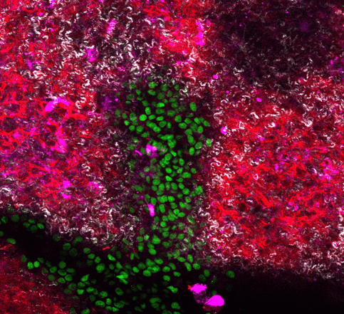
The model, developed in collaboration with clinical scientists from Copenhagen Hospital Region, uses an ECM scaffold from murine lungs, or livers (two frequent sites of metastasis), and challenges cancer cells to colonise and modify the ECM, mimicking the formation of metastatic tumours. The scaffold is housed in a box that provides circulation, nutrition and the possibility to monitor cells as they advance and conquer their new environment. Remarkably, the ECM scaffold induces cells to communicate and behave in ways that imitate tumours in an organism. This opens the possibility to study metastatic disease in a controlled system, performing experiments that cannot be carried out using experimental animals or traditional cell culture systems.
“The key advantage of our model is flexibility of its applications”, said Dr Alejandro Mayorca Guiliani, the designer of the model system, “Metastasis is a threat to cancer patients and a very difficult phenomenon to study. Our model can use scaffolds from different organs and cells carrying different origin and mutations which opens new experimental possibilities to explore the mechanisms of metastasis and other diseases”.
“Using this approach can help to identify and target key factors needed for metastatic tumour formation. It allows us to look at both sides of the process: how cancer cells respond to the ECM and how the ECM is transformed in cancer cells’ favour”, added first author PhD student Maria Rafaeva. One key finding of this study suggests that ECM scaffold can coerce cells to activate an important family of “druggable” proteins, tyrosine kinases, similarly to real tumours.
“Designing effective treatments against metastasis is an urgent task for cancer researchers” said Prof. Janine Erler, senior author of this study, “We hope our model system can become a tool to identify and test new therapeutic strategies against this major clinical problem.”
This work was funded by the European Research Council, Innovation Fund Denmark, Danish Cancer Society, German Cancer Aid, Novo Nordisk Foundation, Lundbeck Foundation, Danish Research Council, among other international agencies.