
In Vivo Imaging (IVIS)
 The system consists of an IVIS detection unit + an isoflurane based anesthesia unit (chamber + IVIS) and is located in the Biocenter animal facility.
All research groups at BRIC and FINSEN can use the system, as well as other groups upon agreement with the facility.
The facility only provides access to the IVIS system and data analysis software (no experiment performed for subparties). Before being granted access to the system, a referent person for the team MUST be trained.
Use of the machine is access based
A referent for each group that wants to use the machine MUST be trained. He/she will be taught how to anesthetise mice, perform a luminescent or fluorescent signal acquisition, analyse the data.
Data analysis
Data are exported from the IVIS computer and can be analysed later on a 4 different computers at BRIC-FINSEN.
Generation of luminescent/fluorescent cell lines
The users can generate their own luminescent/fluorescent cell lines using a lentiviral based system, after checking that the cells are free from any murine pathogens (Felasa tests).
Infection of the cells with these lentiviral particles must be performed in a Felasa clean area of a P2 laboratory, available in BRIC’s P2 laboratory for example.
Online booking system only once people have been trained.
Please contact Yasuko Antoku, Yasuko.Antoku@bric.ku.dk for more details.
For internal UCPH users
- The facility provides reagents necessary to the use of the machine (O2, charcoal cartridges, etc..) on the exception of luciferin (users have to purchase themselves).
- Data analysis on common computers is free of charge.
For external non UCPH users
- Same pricing for internal users.
SNHG5 promotes colorectal cancer cell survival by counteracting STAU1-mediated mRNA destabilization. Damas ND, Marcatti M, Come C, Christensen LL, Nielsen MM, Baumgartner R, et al. Nat Comms. 2016 Dec 22;7:13875.
The hypoxic cancer secretome induces pre-metastatic bone lesions through lysyl oxidase. Cox TR, Rumney RMH, Schoof EM, Perryman L, Høye AM, Agrawal A, et al. Nature. Nature Research; 2015 Jun 4;522(7554):106–10.
Instruments
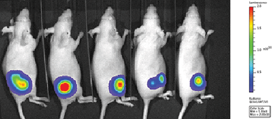
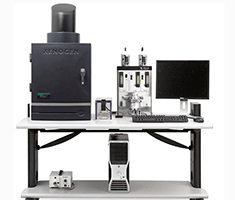 The system consists of an In Vivo Imaging System (IVIS) Lumina II (Perkin Elmer, formerly Xenogen), that can detect luminescent or fluorescent signals from up to 5 mice simultaneously.
For fluorescence, 10 excitation wavelengths (from 430 to 745 nm) and 16 emission filters (from 515 to 875 nm) are available.
Living Image analysis software have been allocated on common computers at BRIC-FINSEN for analysis of the data.
Contact

Yasuko Antoku, staff scientist
Yasuko.Antoku@bric.ku.dk
BRIC - Biotech Research & Innovation Centre
Ole Maaløes Vej 5
DK-2200 Copenhagen
Room 3-4-37